Retinal Imaging
We provide state of the art retinal scans including the following:
-
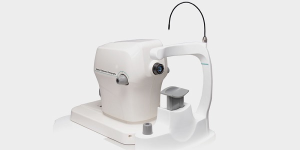
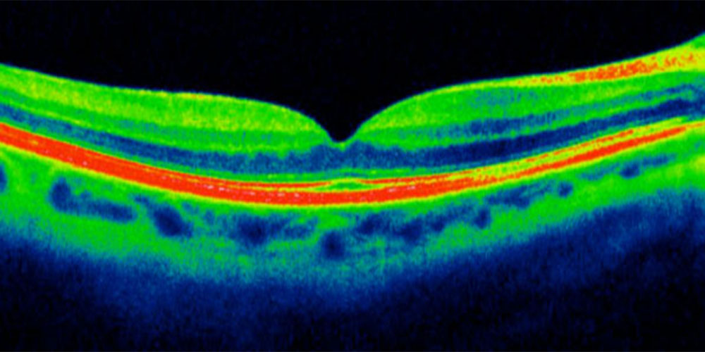
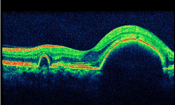
Optical coherence tomography
We have the latest spectral domain OCT scan machine and are able to scan retinal structures with a resolution of 5 microns, giving us very precise information of the disease process. OCT scans are not only used for diagnosis of your retinal condition but also for follow up of diseases on a periodic basis. In the past decade, OCT scans have brought about paradigm shifts in the way we manage retinal diseases. These scans help us plan and deliver the best treatment tailor made for your needs. Patients with the following diseases may require OCT scans periodically:
-
Age related macular degeneration
- Diabetic macular edema
- Retinal Vein occlusion
-
Other conditions that are treated with intraocular injections
-
Age related macular degeneration
-
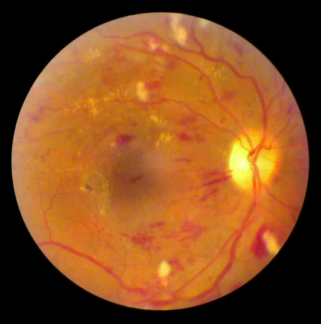
Fundus Photography
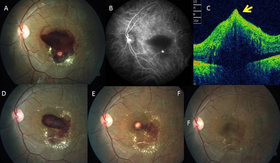
Colour fundus photography helps us capture important information from the fundus such as the exact nature of the problem, size and nature of the pathology and relation of disease with important structural landmarks in the retina. These photos can be saved digitally and sent to you via Email so that you have a permanent record of these findings. The colour fundus photograph is most helpful for follow up and comparison of the disease process over time. Patients with many diseases such as diabetic retinopathy frequently need serial photographs to document changes in the retina over time.
-
Fundus Fluorescein Angiography
We perform angiography of the retina to understand the level of blood flow going through the retinal blood vessels. This helps us to diagnose the exact stage of disease and also plan the correct treatment that will suit you the best. Many diseases such as diabetic retinopathy, retinal vein occlusions and macular degeneration require angiography for this purpose.
